
|
When hunting mammmals, having a basic understanding and visual picture of their anatomy can help you get a clear, clean shot. A shot through the heart or lungs will take the animal down faster, causing them less pain,and ensuring that they don't run away and die someplace you can't find, thereby wasting the meat. A shot through the heart or lungs also ensures that you don't spoil meat by contaminating it with fluid or solids from the digestive tract. All in all a carefully calculated shot with knowledge of the animal's anatomy is of benefit to everyone, the animal included. The following images were created by Dr. Bengt Röken, who worked for 38 years as a Veterinarian at Kolmarden Zoo in Sweden. Dr. Röken used euthanized zoo animals for anatomical studies and dissections. Dr. Röken has published these images previously in hunting magazines to help hunters make clean shots. He has graciously provided some of his work for this project, and though his work is not of caribou but of moose, many hunters who hunt caribou also hunt moose, and there are anatomical similarities. We hope these images will help you visualize the internal anatomy and help with efficient hunting. |
Dr. Röken, in Kenya with giraffes in the background. |
Moose Anatomy Heart and Lung Positioning Depending on Angle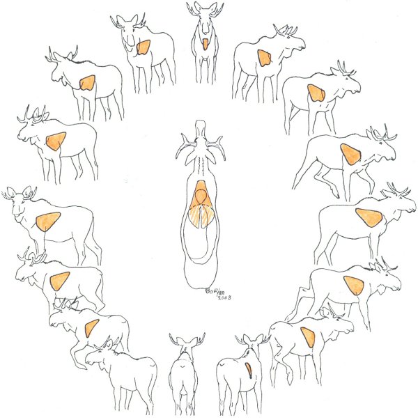
Image © B.O. Roken, DVM, 2008, bengt.roken@gmail.com www.vetroken.se |
Vital organs/shot placement in the moose:Vital organs include the heart, lungs and thoracic spinal column. When heart or both lungs are hit by an expandable bullet, fatal bleeding occurs within minutes. Spinal injury results in immediate collapse and paralysis. If only the upper parts of the lungs are injured a rapid death due to internal bleeding occurs.
|
Drawings of Moose Anatomy - Focusing on the lungs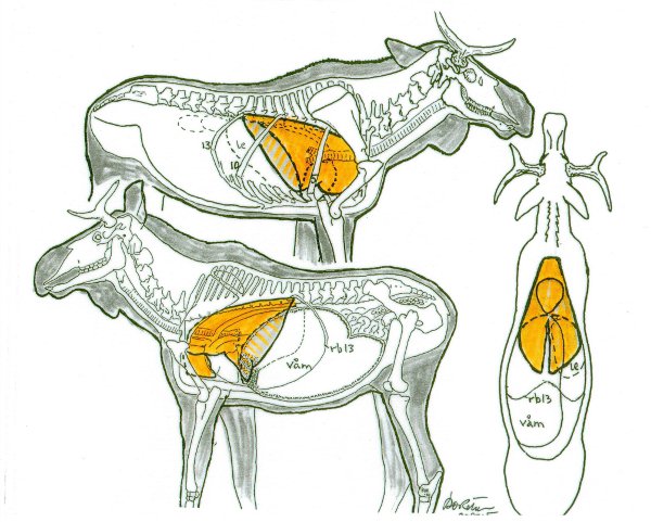
Image © B.O. Roken, DVM, 2001,bengt.roken@gmail.com, www.vetroken.se |
Lungs in front of the diaphragm midline are colored deeper than the parts that lie behind. The right lung is larger than the left, and only the right apical lung lies in front of the heart. |
|
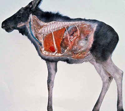
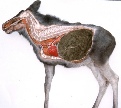
This zoo born moose calf was a small underdeveloped animal, while the muscle mass was weak, and there was minimal fat deposits, the abdominal organs were well developed. During the dissection in a lateral recumbency position, some of the organs were painted. Ribs 1,5,10 and 13 are left in place. The left lung surface is painted orange, the visible part of the heart is painted red and the left jugular vein is painted blue. Image © B.O. Roken, DVM, 1985, bengt.roken@gmail.com, www.vetroken.se |
|
On this dissection, the close proximity between the rumeno-reticulum and thorax and heart, is clearly visible.
Image © B.O. Roken, DVM, 1985, bengt.roken@gmail.com, www.vetroken.se |
|
|
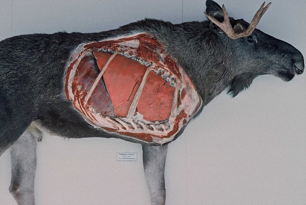
Male moose, 2 years old, prepared September 2000. Image © B.O. Roken, DVM, 1985, bengt.roken@gmail.com, www.vetroken.se |
Copyright © 2011 University of Calgary. Web design by Jesse Invik, jinvik@gmail.com